Bacteria are prokaryotic, unicellular microorganisms, which lack chlorophyll pigments. The cell structure is simpler than that of other organisms as there is no nucleus or membrane bound organelles.
Due to the presence of a rigid cell wall, bacteria maintain a definite shape, though they vary as shape, size and structure.
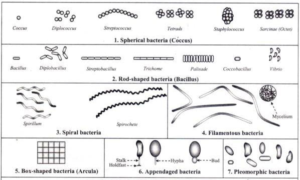
When viewed under light microscope, most bacteria appear in variations of three major shapes: the rod (bacillus), the sphere (coccus) and the spiral type (vibrio). In fact, structure of bacteria has two aspects, arrangement and shape. So far as the arrangement is concerned, it may Paired (diplo), Grape-like clusters (staphylo) or Chains (strepto). In shape they may principally be Rods (bacilli), Spheres (cocci), and Spirals (spirillum).
Size of Bacterial Cell
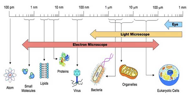
The average diameter of spherical bacteria is 0.5-2.0 µm. For rod-shaped or filamentous bacteria, length is 1-10 µm and diameter is 0.25-1 .0 µm.
- E. coli , a bacillus of about average size is 1.1 to 1.5 µm wide by 2.0 to 6.0 µm long.
- Spirochaetes occasionally reach 500 µm in length and the cyanobacterium
- Oscillatoria is about 7 µm in diameter.
- The bacterium, Epulosiscium fishelsoni , can be seen with the naked eye (600 µm long by 80 µm in diameter).
- One group of bacteria, called the Mycoplasmas, have individuals with size much smaller than these dimensions. They measure about 0.25 µ and are the smallest cells known so far. They were formerly known as pleuropneumonia-like organisms (PPLO).
- Mycoplasma gallicepticum, with a size of approximately 200 to 300 nm are thought to be the world smallest bacteria.
- Thiomargarita namibiensis is world’s largest bacteria, a gram-negative Proteobacterium found in the ocean sediments off the coast of Namibia. Usually it is 0.1—0.3 mm (100—300 µm) across, but bigger cells have been observed up to 0.75 mm (750 µm).
Thus a few bacteria are much larger than the average eukaryotic cell (typical plant and animal cells are around 10 to 50 µm in diameter).
Shape of Bacterial Cell
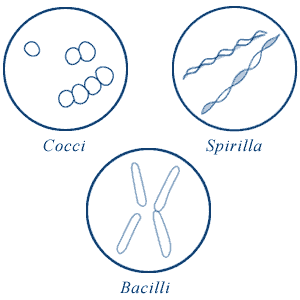
The three basic bacterial shapes are coccus (spherical), bacillus (rod-shaped), and spiral (twisted), however pleomorphic bacteria can assume several shapes.
- Cocci (or coccus for a single cell) are round cells, sometimes slightly flattened when they are adjacent to one another.
- Bacilli (or bacillus for a single cell) are rod-shaped bacteria.
- Spirilla (or spirillum for a single cell) are curved bacteria which can range from a gently curved shape to a corkscrew-like spiral. Many spirilla are rigid and capable of movement. A special group of spirilla known as spirochetes are long, slender, and flexible.
Arrangement of Cocci
Cocci bacteria can exist singly, in pairs (as diplococci ), in groups of four (as tetrads ), in chains (as streptococci ), in clusters (as stapylococci ), or in cubes consisting of eight cells (as sarcinae). Cocci may be oval, elongated, or flattened on one side. Cocci may remain attached after cell division. These group characteristics are often used to help identify certain cocci.
1. Diplococci
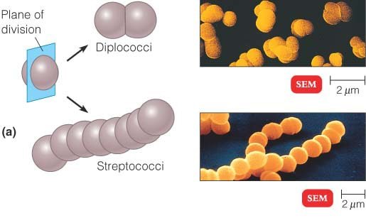
The cocci are arranged in pairs.
Examples: Streptococcus pneumoniae, Moraxella catarrhalis, Neisseria gonorrhoeae, etc.
2. Streptococci
The cocci are arranged in chains, as the cells divide in one plane.
Examples: Streptococcus pyogenes, Streptococcus agalactiae
3. Tetrads
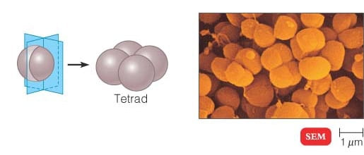
The cocci are arranged in packets of four cells, as the cells divide in two plains.
Examples: Aerococcus, Pediococcus and Tetragenococcus
4. Sarcinae
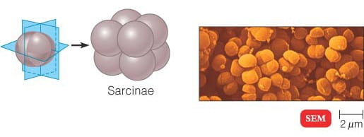
The cocci are arranged in a cuboidal manner, as the cells are formed by regular cell divisions in three planes. Cocci that divide in three planes and remain in groups cube like groups of eight.
Examples: Sarcina ventriculi, Sarcina ureae, etc.
5. Staphylococci

The cocci are arranged in grape-like clusters formed by irregular cell divisions in three plains.
Examples: Staphylococcus aureus
Arrangement of Bacilli
The cylindrical or rod-shaped bacteria are called ‘bacillus’ (plural: bacilli).
1. Diplobacilli
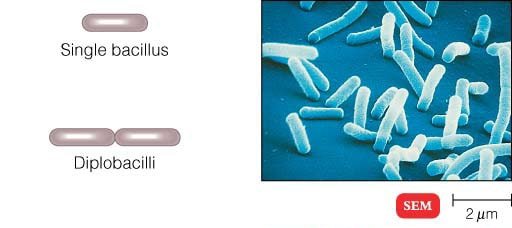
Most bacilli appear as single rods. Diplobacilli appear in pairs after division.
Example of Single Rod: Bacillus cereus
Examples of Diplobacilli: Coxiella burnetii, Moraxella bovis, Klebsiella rhinoscleromatis, etc.
2. Streptobacilli
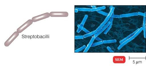
The bacilli are arranged in chains, as the cells divide in one plane.
Examples: Streptobacillus moniliformis
3. Coccobacilli
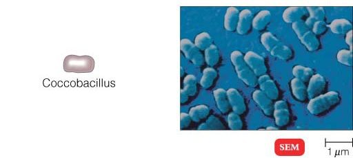
These are so short and stumpy that they appear ovoid. They look like coccus and bacillus.
Examples: Haemophilus influenzae, Gardnerella vaginalis, and Chlamydia trachomatis
4. Palisades
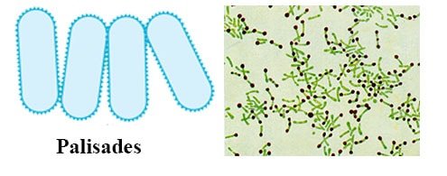
The bacilli bend at the points of division following the cell divisions, resulting in a palisade arrangement resembling a picket fence and angular patterns that look like Chinese letters.
Example: Corynebacterium diphtheriae
Arrangement of Spiral Bacteria
Spirilla (or spirillum for a single cell) are curved bacteria which can range from a gently curved shape to a corkscrew-like spiral. Many spirilla are rigid and capable of movement. A special group of spirilla known as spirochetes are long, slender, and flexible.
1. Vibrio
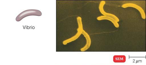
They are comma-shaped bacteria with less than one complete turn or twist in the cell.
Example: Vibrio cholerae
2. Spirilla
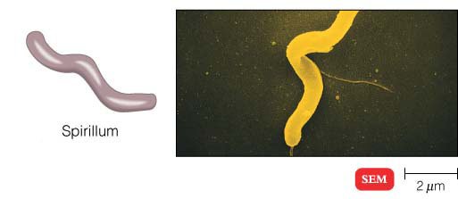
They have rigid spiral structure. Spirillum with many turns can superficially resemble spirochetes. They do not have outer sheath and endoflagella, but have typical bacterial flagella.
Example: Campylobacter jejuni, Helicobacter pylori, Spirillum winogradskyi, etc.
3. Spirochetes
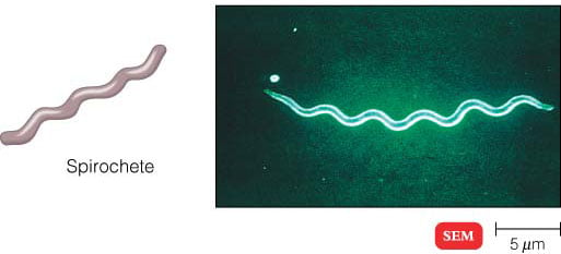
Spirochetes have a helical shape and flexible bodies. Spirochetes move by means of axial filaments, which look like flagella contained beneath a flexible external sheath but lack typical bacterial flagella.
Examples: Leptospira species (Leptospira interrogans), Treponema pallidum, Borrelia recurrentis, etc.
Others Shapes and Arrangements of Bacteria
1. Filamentous Bacteria
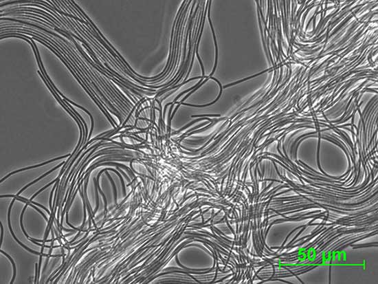
They are very long thin filament-shaped bacteria. Some of them form branching filaments resulting in a network of filaments called ‘mycelium’.
Example: Candidatus Savagella
2. Star Shaped Bacteria
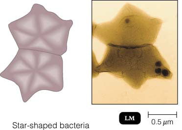
Example: Stella
3. Rectangular Bacteria
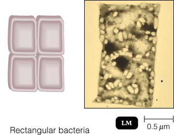
Examples: Haloarcula spp (H. vallismortis, H. marismortui)
4. Pleomorphic Bacteria
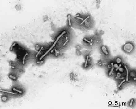
These bacteria do not have any characteristic shape unlike all others described above. They can change their shape. In pure cultures, they can be observed to have different shapes.
Examples: Mycoplasma pneumoniae, M. genitalium, etc.

How do I know when the bacteria moves unilaterally or in pairs?
How to figure the diffrence between them?
To answer Shahanaz akter’s question….
In the classification of bacteria by staining, differential staining methods (gram stain and acid fast stain methods) are used . In the gram staining method bacteria are stained as either positive or negative gram cells using a dye-iodine complex which permeat the cell wall of a gram- negative cell but not gram positive bacteria cell as it permit the outflow of the iodine complex during decoloration..
As in acid- fast staining certain bacteria such as the tubercle baccilli resist decoloration with acids. This is how bacteria are classified by staining method.
As for oxygen based classification of bacteria, depending on the influence of on growth and viability, bacteria are divided into aerobic and anaerobes
Aerobic bacteria require oxygen for growth while anaerobes bacteria don’t require bacteria for growth.
You said in the introduction that bacteria do not have chlorophyll, but that is untrue. Bacteria invented chlorophyll* about 3 billion years ago, and one of those bacteria went on to become the ancestor of today’s chloroplasts. There are still many kinds of free-living photosynthetic bacteria, or cyanobacteria, in existence.
* Unless you are not counting bacteriochlorophyll as chlorophyll? But that would be both pedantic and confusing to your readers…
Why coccus is in the shape of cocci ?and rod is in the shape of rod ? What’s the principle it has ?
Hi, Sheshu, I’m no expert, but I do know in biomaterials engineering that nano-sized particles that are rod shaped are much more reactive in the body than other shapes. For example, if you have gold nano particles that are round in shape, and you introduce them into the body for certain types of medical therapies, the nano particles are inert. The body does not react to them. But when you introduce rod-shaped gold nano particles into the body, the body reacts quickly and severely. The rod shape is essentially getting rejected by the body. I hope that gives you some idea as to how shape can change a bacteria’s action in our bodies.
what about classification of bacteria based on staining and ased on oxygen requirement??
*very you immigrant
Nice listing, although there is a serious typo in the section on the size of bacteria. You state that Epulosisicum fishelsoni has a certain size. First, the genus name is Epulopiscium. The “piscium” part comes from the fact that they live inside of fish. Second, and more egregious, is that you state the length to be 600mm. That is about 2/3 of a meter, which is longer than most of the fish they inhabit. I think your units here should be micro-meters not milli-meters.
Cheers
Thank you so much for the correction. I corrected it now 🙂
Cheerss
Gram +/_ bacteria identification flow chart with photos ??? A method which makes it easy to remember
Thiomargarita namibiensis is a gram-negative coccoid Proteobacterium, found in the ocean sediments of the continental shelf of Namibia. It is the largest bacterium ever discovered, as a rule 0.1–0.3 mm (100–300 µm) in diameter, but sometimes attaining 0.75 mm (750 µm). Cells of Thiomargarita namibiensis are large enough to be visible to the naked eye. Although the species holds the record for the most massive bacterium, Epulopiscium fishelsoni – previously discovered in the gut of surgeonfish – grows slightly longer, but narrower.
your notes are really of great help. I wish you were my supervisor or lecturer. please I would like to know if you have a writeup on CLEF agar.
thanks
Very interesting information. But what is Steptobacilli? Is that also part of human microflora, is it pathogenic??
its a pathogenic