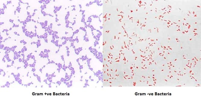
Gram Staining is the common, important, and most used differential staining techniques in microbiology, which was introduced by Danish Bacteriologist Hans Christian Gram in 1884. This test differentiate the bacteria into Gram Positive and Gram Negative Bacteria, which helps in the classification and differentiations of microorganisms.
Principle of Gram Staining
When the bacteria is stained with primary stain Crystal Violet and fixed by the mordant, some of the bacteria are able to retain the primary stain and some are decolorized by alcohol. The cell walls of gram positive bacteria have a thick layer of protein-sugar complexes called peptidoglycan and lipid content is low. Decolorizing the cell causes this thick cell wall to dehydrate and shrink, which closes the pores in the cell wall and prevents the stain from exiting the cell. So the ethanol cannot remove the Crystal Violet-Iodine complex that is bound to the thick layer of peptidoglycan of gram positive bacteria and appears blue or purple in color.
In case of gram negative bacteria, cell wall also takes up the CV-Iodine complex but due to the thin layer of peptidoglycan and thick outer layer which is formed of lipids, CV-Iodine complex gets washed off. When they are exposed to alcohol, decolorizer dissolves the lipids in the cell walls, which allows the crystal violet-iodine complex to leach out of the cells. Then when again stained with safranin, they take the stain and appears red in color.
Reagents Used in Gram Staining
- Crystal Violet, the primary stain
- Iodine, the mordant
- A decolorizer made of acetone and alcohol (95%)
- Safranin, the counterstain
Procedure of Gram Staining
- Take a clean, grease free slide.
- Prepare the smear of suspension on the clean slide with a loopful of sample.
- Air dry and heat fix
- Crystal Violet was poured and kept for about 30 seconds to 1 minutes and rinse with water.
- Flood the gram’s iodine for 1 minute and wash with water.
- Then ,wash with 95% alcohol or acetone for about 10-20 seconds and rinse with water.
- Add safranin for about 1 minute and wash with water.
- Air dry, Blot dry and Observe under Microscope.
Interpretation
Gram Positive: Blue/Purple Color
Gram Negative: Red Color

Examples
Gram Positive Bacteria: Actinomyces, Bacillus, Clostridium, Corynebacterium, Enterococcus, Gardnerella, Lactobacillus, Listeria, Mycoplasma, Nocardia, Staphylococcus, Streptococcus, Streptomyces ,etc.
Gram Negative Bacteria: Escherichia coli (E. coli), Salmonella, Shigella, and other Enterobacteriaceae, Pseudomonas,Moraxella, Helicobacter, Stenotrophomonas, Bdellovibrio, acetic acid bacteria, Legionella etc
Animation
Download Animation from Below:
Gram Staining Animation

Is it only gram staining techniques that can be used to differentiate bacteria?
Gram positive bacteria have thick cell wall peptidoglycan in their cell wall which will make it to retain the complex of crystal violet and iodine when decolorized by acid which will make it to appear as blue or purple. while gram negative bacteria have thin cell wall peptidoglycan when decolorized by an acid, the complex removed due to it’s cell wall and it takes the color of safranin.
What are the gram reaction and staining of various organisms??
Gram positive turns blue
Gram negative turns pink
Why circle drawn under the smear using pencil?
That circle indicates or represents naming: Naming the slide by the owners of the sample under analysis to avoid mistakes. Naming of a slide in every analysis or staining process is inevitable.
why is a gram staining classified as a differential staining?
what is the significant about gram staining?
it mechanism of action for gram staining?
There is a use of two stains to make difference
It helps know gram negative and gram positive bacterials based on the cell wall
Cell wall composition
The cell wall has peptidoglycan which helps with their identification. Gram positive have a thicker layer of peptidoglycan compared to gram negative. That is why the the gram +ve is able to retain the primary stain
Gram staining is a differential stain because it stains bacteria as gram positive or gram negative.
Gram staining is important because it helps us differentiate different classes of bacteria as either gram positive or gram negative.
It is also important as it helps distinguish between the bacteria and the background of the microscope
Is lugol’s Iodine used as a mordant in gram staining
Yes, of course.
Is it possible to re-stain a slide? What is the procedure?
is there other alternative beside gram stain which are used to clasify bacteria
AFB staining method you can look it up
Why is acid fast stain more prefereble than gram stain ?
Because it involves heating which easily breaks the fatty substances in the cell wall of the bacteria.
Afb is used in staining mycobacterium
Can gram stain done on direct urine? (II mean before culture)
please what is the underlying principles behind some bacteria being gram negative and gram positive?
Due to composition of cell wall
The principle is about having thin or thick peptidoglycan that can either be removed or not during staining hence some retain purple colour.
gram positive Bacteria have a thick cell wall of peptydoglycan and gram negative Bacteria have thin peptydoglycan cell wall and a lipid layer also present in gram negative bacteria hence after all steps of procedure of gram staining, gram positive bacteria will remain coloured purple or bluish due to thick cell wall and gram negative bacteria will not able to stain and when we wash ot with water and decolorizer then stain will wash out , not absorbed by the cell wall of gram negative bacteria hence it will appear in only pink color due to counter stain .
i hope it will helpful to you
Is because of their cell wall composition.
The thin and thick peptidoglyca cell wall
What will be the final color of Gram (+) bacteria if mordant will be omitted?
Purple colour
the reason why mordant is use is to grow the microorganism
A mordant fixes or binds cells intensifying stains in cell or tissue to allow for visual clarity under a microscope.
Pls wat will happen when decolouriser is left out
Decolizer is needed for differencites ….so why it left out
If decolorizer is left out, it won’t dissolve the lipids in the cell walls, and therefore, the staining process would be incomplete and it won’t allow the cells to be stained.
Can safranin be called a differential stain
I would say yes. It can be considered a differential stain because its used to better differentiate between microorganisms and/or structures while viewed under a microscope.
What does iodine do In staining ? What happen if we don’t use iodine ?
The role of iodine is that it act as grams mordant and increases the interaction between bacterial cell wall and the dye (crystal violet) so that the dye is more tightly bound on the cell and is more stained.
It forms di-iodine complex,If the bacteria is negative then it broken by OH group of alcohol
Otherwise it will not broken by OH group of alcohol then the bacteria is gram negative
Don’t you think there should be another staining similar to gram staining
Even dilute cabolfuchsin is another counter stain that (1:9)dilution
Hi
Very much appreciated
Would be great if u also add some points about the modifications and variations and also gram variable bacteria and some general info about why gram staining is needed and where it is not indicated !!
Because in medicine what accompanies practicals are viva and these questions can be very important
Cheers
Gud luck
Greatly impressed by this article. Painstakingly put together in a simple language. I studied microbiology at Polytechnic(HND) and presently a 300 level student of Lead City University,Ibadan, Nigeria studying Biology Education.This piece has really helped in solving my Assignment on Biological Techniques(BIO 315)
All enterobcteriaceae are gram negative bacteria but not all gram negative are enterobcteriaceae to distinguish the enterobcteriaceae you have to do oxidase test because enterobcteriaceae are oxidase negative bacteria
Great!
But if i may ask, which step in Gram staining tech can be omited without affecting the final result?
Mr Len artifacts results from poor methods of rinsing of slides.
heat fixing of smear blackened and disolve the lipoid cpnt of the cell as well as it schring of cell cpnt.
Beside, what is the color of safranin?
Try fixing with 100% Methanol for one minute and letting the slide dry, rather than heat fixing..preserves cellular integrity and reduces artifact caused by heat fixation. Good for for CSF’s, pus and epithelial cells. Also, use acetone alcohol – 50% acetone and 50% of 95% ETOH to decolorize. More of a controlled decolorization.
Len Fligg, ASCP cm
If Gram positive organisms have such complex cell wall that could defy decolorisation,why then is a mordant used?
the function of the mordant is to fix (attach) the crystal violet to the cell wall (peptidoglycan cell wall)so as not to be washed away easily thereby forming a complex as stated earlier. Moreover, the test procedure is performed to differentiate between the two groups of bacteria.
What is other type of counter stain ?
And what about over decolorization
Neutral red another type of counter stain