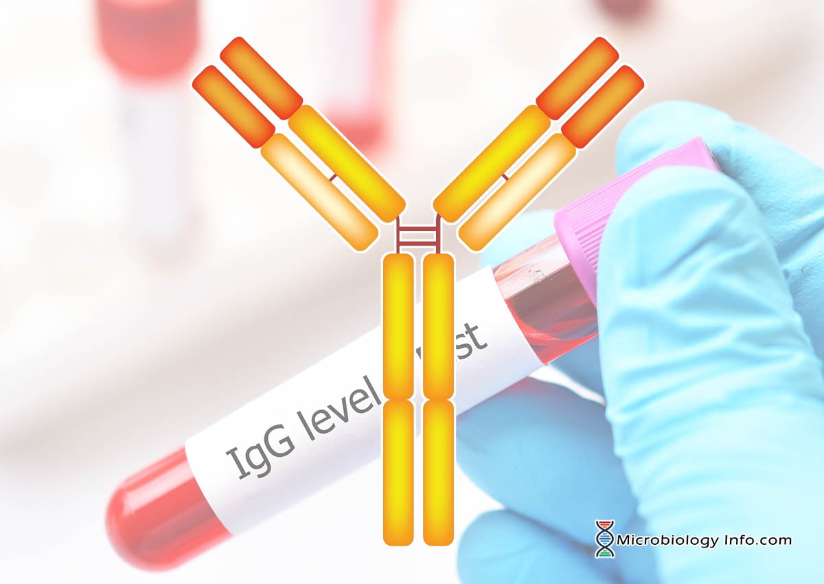
Properties of Immunoglobulin G (IgG)
- Immunoglobulin G (IgG) is the predominant glycoprotein present in human serum, constituting about 75-80% of total immunoglobulin in humans which accounts for 10 to 16mg/ml serum concentration and is produced as part of the secondary immune response to an antigen.
- IgG is usually found as monomers and has an average half-life of 23 days and is the only isotype that crosses the placenta.
- Unlike IgM molecule, the IgG molecule is small and is able to penetrate extracellular and intracellular spaces and sIgG antibodies are distributed equally between the intra- and extravascular pools.
- IgG binds antigens with both high affinity and low avidity.
- In humans, there are four sub-isotypes of heavy chains, 1, 2, 3, and 4 with the corresponding formation of the four subclasses of IgG: IgG1, IgG2, IgG3, and IgG4 numbered by their amount present in the serum and also with differences based on the antigenic differences in the structure of the heavy chain. In mice, the four chain sub isotypes are 1, 2, 2, and 3, and the corresponding subclasses of mouse IgG antibodies are IgG1, IgG2a, IgG2b, and IgG3, respectively.
- The small differences present in the amino acid sequence and disulfide bonding between subclasses confer small differences in physical properties, which in turn are associated with variable longevity in the serum and differences in effector functions of the isotypes.
- All IgG subclasses are found at significant levels at the mucosal barriers. Mucosal IgG reaches the mucosal surface either being produced locally by mucosal plasma cells or systemically by the neonatal Fc Receptor (FcRn).
Structure of Immunoglobulin G (IgG)
- Immunoglobulin G (IgG) is a large glycoprotein of 150KDa molecular weight existing in monomeric configurations.
- IgG are glycoproteins formed of four polypeptide chains, consisting of two identical copies of each of two kinds of polypeptide chain: light chain (κ or λ) and heavy chain gamma (γ) linked together by disulfide bonds and non-covalent forces.
- Its H-chain type is gamma (γ heavy chains) about 50kDa molecular weight and the light chain can be either Kappa (κ) or Lambda (λ) type of about 25 kDa molecular weight.
- The heavy chains are linked to each other by disulphide bonds and each heavy chain is linked to a light chain by disulphide bonds. The resulting tetramer has two identical halves, which together form the Y-like shape.
- The light chain comprises of two domains. The first domain is Light chain Variable domain (VL) which confers variability in the sequence of the light chain. The other domain is known as the Light chain Constant domain (CL).
- The heavy chain is made up of one Variable domain (VH) and three Constant domains (CH1, CH2 and CH3) along with hinge region.
- The VL and VH fold together to make up the Variable region of the IgG molecule and confer on it its antigen binding specificity.
- The VL and VH and CL and CH1 domains fold together to make up the Fab or antibody binding fragment of the IgG molecule.
- The CH2 and CH3 domains of the two heavy chains fold together to form the Fc or crystallizable fragment of the molecule which bears a highly conserved N-glycosylation site.
- The hinge region is rich in proline residues, rendering flexibility to the molecule and as a consequence, the two antigen-binding arms of IgG antibodies can assume a wide variety of angles with respect to one another, which in turn facilitates efficient antigen binding.
- In addition to these proline residues present, the hinge region also contains a number of cysteines residues, which participate in heavy-chain dimerization.
Subclasses of Immunoglobulin G (IgG)
There are four subclasses of IgG: IgG1, IgG2, IgG3, and IgG4 which are differentiated on the position of interchain disulfide bonds, basis of the size of the hinge region and molecular weight along with differences based on the antigenic differences in structure of the heavy chain.
IgG1
- IgG1 comprises 60 to 65% of the total main subclass of IgG and is usually produced in response to protein antigens which are T-dependent antigens.
- The molecular weight of IgG1 is 150kDa.
- The IgG1 are particularly good opsonins and mediators of ADCC because FcγRs bind sIgG1 antibodies with high affinity.
- IgG1 binds to the Fc-receptor of phagocytic cells and can activate the complement cascade via binding to the C1 complex. However IgG1 is somewhat less efficient in complement fixing.
- IgG1 immune response can already be measured in newborns and reaches its typical concentration in infancy.
- A deficiency in IgG1 isotype is typically a sign of hypogammaglobulinemia.
IgG2
- IgG2 is the second largest of IgG isotypes which comprises 20 to 25% of the main subclass, usually produced in response to carbohydrate/polysaccharide antigens which are T-independent antigens.
- FcγRs hardly bind to sIgG2.
- IgG2 crosses the placenta with much lower efficiency.
- IgG2 is even less efficient in complement fixing as compared to IgG1.
- IgG2 is especially important as a repository for antibodies against antigens from many common bacteria that cause pneumonia.
- A deficiency in IgG2 is the most common among all IgG isotype deficiencies and is associated with recurring airway/respiratory infections in infants.
IgG3
- IgG3 comprises around 5 to 10% of total IgG and has a molecular weight of 170 kDa.
- They play a major role in the immune responses against protein or polypeptide antigens which are T dependent antigens.
- The IgG3 are particularly good opsonins and mediators of ADCC because FcγRs bind sIgG3 antibodies with high affinity.
- IgG3 has an extended hinge in the CH2 domain of the Ig structure.
- IgG3 is the most efficient complement-fixing IgG subclass.
IgG4
- IgG4 comprises usually less than 4% of total IgG.
- Carbohydrate antigens elicit the production of IgG4.
- IgG4 is unable to bind C1q and so cannot fix complement at all in the classical pathway.
- IgG4 has a very short hinge region.
- FcγRs generally bind less well to sIgG4 as compared with sIgG1 and sIgG3.
- In the past, testing for IgG4 has been associated with food allergies, and recent studies have shown that elevated serum levels of IgG4 are found in patients suffering from cholangitis, sclerosing pancreatitis and interstitial pneumonia caused by infiltrating IgG4-positive plasma cells. The exact role of IgG4 is still mostly unknown.
Functions of Immunoglobulin G (IgG)
- The IgG antibodies are the main line of acquired defense and the main potentiator of a body’s specific humoral response to pathogens in the host extracellular fluids, including blood, lymph and saliva.
- An important function of IgG in the lower respiratory tract is its specific antibody activity against microbial agents or antigens. IgG antibody in the alveolar spaces with specific activity or affinity for a microbe could opsonize it and then interact with the complement cascade to create a lytic situation that might kill the microbe directly, or the opsonized microbe could be phagocytized by an alveolar macrophage and killed by the phenomenon called phagocytosis.
- Only IgG antibodies can cross the mammalian placenta (with the exception of IgG2), and maternal IgG1, IgG3, and IgG4 molecules have been found to be crucial for conferring the passive immunity that protects the developing human fetus and newborn in the first few months of life.
- The IgG antibodies render several effector functions including the neutralization of pathogen (neutralizing many bacterial endotoxins and viruses) by coating the pathogen and occupying its binding sites so that it cannot affect cells, opsonizing microbes by marking it for phagocytosis and killing and the activation of complement cascade to lyse pathogen cells.
- IgG is produced in a delayed response to an infection and can be retained in the body for a longer period of time. The longevity of the IgG molecule in serum makes it most useful for passive immunization by transfer of this antibody. Detection of IgG usually indicates vaccination or a prior infection.
References
- Reynolds H.Y. 1998. Immunoglobulin G and Its Function in the Human Respiratory Tract. Volume 63, Issue 2, Pages 161-174.
- Gocki, J., & Bartuzi, Z. (2016). Role of immunoglobulin G antibodies in diagnosis of food allergy. Postepy dermatologii i alergologii, 33(4), 253-6.
- Abbas A.K and Lichtman A.H. Cellular and molecular immunology. Fifth edition. Page no.34-43.
- Owen, J. A., Punt, J., & Stranford, S. A and Jones, P.P (2013). Kuby Immunology (7 ed.). New York: W.H. Freeman and Company.
- Delves, P.J., Martin, S.J., Burton, D.R. and Roitt, I.M. (2011). Roitt’s Essentail Immunology. 12th edition. A John Wiley and Son’s, Ltd, Publication.
- Cruse, J.M. and Lewis, R.E. (2010). Atlas of Immunology. Third edition. CRC Press. Taylor and Francis Group, 6000 Broken Sound Parkway NW, Suite 300, Boca Raton, FL 33487-2742.

