Phosphatidylcholine or simply, lecithin is a substance widely distributed in animal tissues, egg yolk, and some higher plants, consisting of phospholipids linked to choline.
Some microorganisms possess lecithinase, also called phospholipase C, which is an enzyme that splits the phospholipid lecithin. Such lecithinase activity is used to characterize several gram positive and gram negative bacteria.
Objectives
- To determine the ability of microorganisms to produce the enzyme lecithinase.
- To identify the bacteria on the basis of its lecithinase activity
Principle
Egg Yolk Agar is a differential and enriched medium used in the isolation and presumptive differentiation of different species based on their lecithinase activity. Lecithin is a normal component of the egg yolk. In egg yolk agar, the lipoprotein component lecithovitellin can be split by lecithinase into phosphorylcholine and an insoluble diglyceride, which results in the formation of a precipitate in the medium.
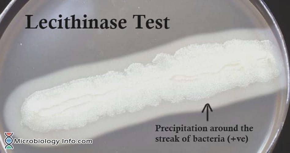
Microorganisms that possess the enzyme lecithinase break down lecithin to insoluble diglyceride and phosphorylcholine, which results in a white opaque zone of precipitation that spreads beyond the edge of the colony. Such an opaque halo, surrounding the colony when grown on the egg yolk agar medium indicates positive lecithinase activity of the test organism.
Media: Egg Yolk Agar
Composition:
Pancreatic Digest of Casein 15.0gm, Vitamin K 1 10.0gm , Sodium Chloride 5.0gm, Papaic Digest of Soybean Meal 5.0gm, Yeast Extract 5.0gm, L-Cystine 0.4gm, Hemin 5.0gm, Egg Yolk Emulsion 100.0ml, Agar 20.0gm
Final pH 7.0 +/- 0.3 at 25ºC.
Procedure
- Take a loopful of test organism and streak it as a straight line on the plate.
- Incubate anaerobically in a gas pak jar immediately after streaking and transfer into the incubator maintained at 35-37oC for 24-48 hours for anaerobes and for aerobes incubate the plate at 35-37oC for 24-48 hours.
- Examine the plate for the opalescent halo surrounding the inoculums.
Results
- Positive test:Appearance of a white, opaque, diffuse zone that extends into the medium surrounding the colonies.
- Negative test:Absence of a white, opaque zone extending from the edge of the colony.
Uses
- Bacterial lecithinases are of special interest because of the possible role of these enzymes in pathogenicity.
- In identification of Clostridium perfringens, Staphylococcus aureus, Pseudomonas aeruginosa or Listeria monocytogenes .
- The lecithinase activity of S. aureus is used in detection of coagulase-positive strains, because of high link between lecithinase activity and coagulase activity.
- Can be used to differentiate between certain species within the genus Bacillus.
Limitations
- It is recommended that biochemical, immunological, molecular, or mass spectrometry testing be performed on colonies from pure culture for complete identification.
- Maintenance of anaerobic condition is compulsory.
- A negative lecithinase test should be compared to an un-inoculated control plate, as lecithinase can diffuse throughout the entire agar plate and make interpretation difficult.
References
- Tille, P. M., & Forbes, B. A. (2014). Bailey & Scott’s diagnostic microbiology (Thirteenth edition.). St. Louis, Missouri: Elsevier.
- Cappuccino J.G. and Sherman N. 2008. Microbiology: A Laboratory Manual, 8th ed. Pearson Benjamin Cummings, San Francisco, CA, USA.
- https://catalog.hardydiagnostics.com/cp_prod/Content/hugo/EggYolkAgarModified.html
- www.vetbact.org/popup/popup.php?id=34&LANG=en
- https://www.ncbi.nlm.nih.gov/pmc/articles/PMC291196/?page=1
- vlab.amrita.edu/?sub=3&brch=73&sim=974&cnt=1