Immunoglobulin D (IgD) is a unique immunoglobulin discovered in 1965.
IgD is found in very low concentrations in serum, far below those of IgG, IgA, and IgM, but much higher than that of IgE, and does not activate the complement pathway.
IgD (also called as Membrane-bound IgD) is the major antigen receptor isotype on the surfaces of mature, peripheral B cells that already express mIgM but a subset of IgM–IgD+ B cells has been found in the upper respiratory mucosa.
IgD is, therefore, the second Ig isotype to be synthesized by a B cell, and first appears on its surface early in B cell development.
IgD starts to be expressed when the B cell exits the bone marrow to populate peripheral lymphoid tissues. When a B cell reaches its mature state, it co-expresses both IgM and IgD on its surface.
The heavy chains (δ) of IgD from the surface of B cells are covalently linked by only one disulfide bridge, appearing close to the carboxy terminus which allows a higher degree of freedom of the antigen-binding sites than for the other immunoglobulin isotypes.
Serum IgD and membrane-bound IgD are similar antigenically however they differ in susceptibility to proteolysis by plasmin.
The majority of the cell-bound IgDs are of the κ type, while 90% of the monoclonal IgDs in serum are of the λ type. In IgD-secreting cells, the IgD molecule found is the preferential association between δ heavy chains and λ light chains.
Structure of Immunoglobulin D (IgD)
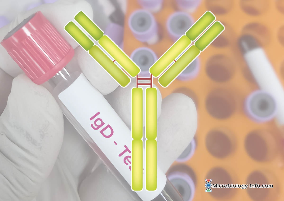
IgD is monomeric in nature and richly glycosylated with a molecular weight of 180KDa.
Because it has a half-life of 2 to 3 days, the plasma concentration is less than 1% of the total immunoglobulin in serum which accounts for 0-0.4 mg/ml.
IgD is composed of two delta (δ) heavy chains with 62KDa molecular weight and two light (κ or λ) chains with 23 KDa molecular weight.
Also, in addition to the disulfide bonds linking the chains together, there are intrachain disulfide links that divide each chain into areas called domains.
The light chains have two domains, one constant and one variable.
The heavy chains have four domains, one variable and three constant-region domains.
The hinge (H) region of IgD is diverse in terms of length, amino acid composition and glycosylation.
IgD has a long hinge region consisting of an amino-terminal region rich in alanine and threonine residues and a carboxy-terminal region rich in charged lysine, glutamate and arginine residues, with up to seven O-linked glycans.
The long “hinge” region between the Fab and Fc region renders the IgD molecule very susceptible to proteolytic degradation with the production of Fc and Fab fragments.
In addition, it also renders the IgD molecule capable of acquiring a flexible T shape rather than the traditional Y shape of other antibody isotypes, with two antigen-binding Fab arms swiveling at the two sides of the Fc region.
On the other side, the flexibility of the T shape may help IgD to bind epitopes that have a low density on the surface of particulate antigens and also renders the molecule flexible, thus enhancing antigen binding.
IgD is bound to B cells via the Fc receptor or is free in serum.
Functions of Immunoglobulin D (IgD)
The precise function of mIgD is still unknown, but it is capable of sending signals to the B cell nucleus via its associated Igα/Igβ heterodimer.
It is thus possible that mIgD either regulates B cell maturation or lengthens the life span of mature B cells in the periphery. IgD can also participate in the generation and maintenance of B-cell memory and might have an important role in the transition from a stage of susceptibility to induction of B-cell tolerance to one of responsiveness.
The IgD molecule functions primarily as an antigen receptor on B cells and is probably involved in regulating B cell function when it encounters antigen.
The inherent flexibility of mIgD due to the long hinge region in the structure may allow the molecule to bind to antigens featuring epitopes that are widely spaced and cannot be bound by the more rigid mIgM molecule.
IgD enhances humoral immune responses through its induction of IgD-receptor expression on T lymphocytes. When IgM and IgD expressed on a B cell surface interact with an antigen for which they are specific, the antigen is internalized, and processed and presented to helper T cells which activate the B cells to proliferate and differentiate into plasma cells, as a result initiate the development of a humoral immune response.
IgD can have a regulatory role such as enhancing a protective antibody response of the IgM, IgG, or IgA isotype, or interfering with viral replication.
Secreted IgD is found to enhance mucosal immunity because of the abundance of IgM−IgD+ B cells in the upper respiratory mucosa and also secreted IgD binds microbial virulence factors as well as pathogenic respiratory bacteria and viruses.
Secreted IgD plays an elusive function in mucosal secretions, blood and on the surface of innate immune effector cells such as basophils.
Some scientists believe that IgD may have some role in allergic reactions. Although the nature of the IgD receptor remains elusive, cross-linking of IgD on basophils tends to stimulate the release of immunoactivating, proinflammatory and antimicrobial mediators.
References
Abbas A.K and Lichtman A.H. Cellular and molecular immunology. Fifth edition.
Goldsby, R.A., Kindt, T.J., Osborne, B.A. and Kuby J. Kuby Immunology. Fifth edition.
Owen, J. A., Punt, J., & Stranford, S. A and Jones, P.P (2013). Kuby Immunology (7 ed.). New York: W.H. Freeman and Company.
Delves, P.J., Martin, S.J., Burton, D.R. and Roitt, I.M. (2011). Roitt’s Essentail Immunology. 12th edition. A John Wiley and Son’s, Ltd, Publication.
Cruse, J.M. and Lewis, R.E. (2010). Atlas of Immunology. Third edition. CRC Press. Taylor and Francis Group, 6000 Broken Sound Parkway NW, Suite 300, Boca Raton, FL 33487-2742.
Chen, K., & Cerutti, A. (2011). The function and regulation of immunoglobulin D. Current opinion in immunology, 23(3), 345-52.
Vladutiu A.O. (2000). Immunoglobulin D: Properties, Measurement, and Clinical Relevance. Clinical and Diagnostic Laboratory Immunology. doi: 10.1128/CDLI.7.2.131-140.