It has been reported that the enzyme β-glucuronidase is present in most strains of E. coli (97%).Organisms other than E. coli (e.g., Salmonella, Shigella, Staphylococcus, Streptococcus, etc.) also possess the enzyme β-glucuronidase. Hence, the detection of the β-glucuronidase enzyme is commonly employed in laboratories to identify and differentiate such organisms.
The substrate, 4-methylumbelliferyl-β-D-glucuronide (MUG), is both sensitive and selective for detection of β-glucuronidase activity. Hence, the MUG test in conjunction with oxidase, indole, and lactose fermentation can be performed to effectively identify E. coli and related organisms.
Objective
To identify glucuronidase activity in orgnasims by fluorescence, under a long-wave (365nm) UV light source.
Principle
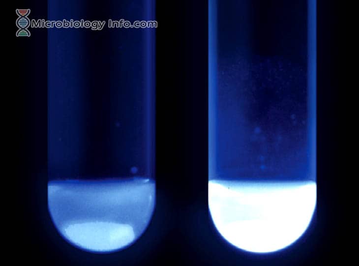
E. coli and other Enterobacteriaceae produce the enzyme β-d-glucuronidase, which hydrolyzes β-d-glucopyranosid-uronic derivatives to aglycons and d-glucuronic acid. The substrate 4-methylumbelliferyl-β-d-glucuronide is impregnated into the disk and is hydrolyzed by the enzyme to yield the 4-methylumbelliferyl moiety, which fluoresces blue under long wavelength ultraviolet light. Hence, if the test organism produces the enzyme glucuronidase it will break down the substrate which will ultimately result in a fluorescence indicating a positive test. Incase of the absence of desired enzyme activity, the substrate is not broken down resulting in no fluorescence on test, which is a negative test.
Media:
MUG Disks are prepared by impregnating MUG (4-methylumbelliferyl-ß-D-glucuronide) onto a high quality filter paper disk.
Method
- Wet the disk with 1 drop of water. Do no saturate.
- Using a wooden applicator stick, rub a portion of a colony from an 18- to 24-hour-old pure culture onto the disk.
- Incubate at 35°-37°C in a closed container for up to 2 hours.
- Observe disk using a 366-nm ultraviolet light.
Expected Results
- Positive: Electric blue fluorescence
- Negative: Lack of fluorescence
Uses
- The test is used to presumptively identify various genera of Enterobacteriaceae.
- To characterize verotoxin-producing Escherichia coli. (verotoxinproducing strains of E. coli do not produce MUG, and a negative test result may indicate the presence of a clinically important strain.)
- It aids in the detection of Escherichia colifrom water and food samples.
Limitations
- Colonies isolated from media containing dyes (EMB, MAC), should not be tested because it may make the interpretation difficult.
- The test can be reliable if performed on oxidase-positive organisms only, because some oxidase-negative organisms naturally fluoresce.
- It is recommended that biochemical, immunological, molecular, or mass spectrometry testing be performed on colonies from pure culture for complete identification.
- coli 0157:H7 will be MUG negative.
- Some strains of Shigella are MUG-positive. Serological testing may be required to differentiate Shigella and E. coli.
References
- Tille, P. M., & Forbes, B. A. (2014). Bailey & Scott’s diagnostic microbiology (Thirteenth edition.). St. Louis, Missouri: Elsevier.
- https://catalog.hardydiagnostics.com/cp_prod/Content/hugo/MUGDisks.html
- https://eportal.mountsinai.ca/Microbiology//manual/tech/tech25.pdf
- www.himedialabs.com/TD/M1461.pdf
- https://www.atcc.org/en/Global/FAQs/D/4/E_coli_and_the_MUG_test-816.aspx
- https://assets.thermofisher.com/TFS-Assets/LSG/manuals/IFU21135.pdf
Similar Posts:
- Salmonella Shigella (SS) Agar- Composition, Principle, Uses, Preparation and Result Interpretation
- PYR Test- Principle, Uses, Procedure and Result Interpretation
- ONPG Test – Principle, Procedure, Uses and Interpretation
- Oxidase Test- Principle, Uses, Procedure, Types, Result Interpretation, Examples and Limitations